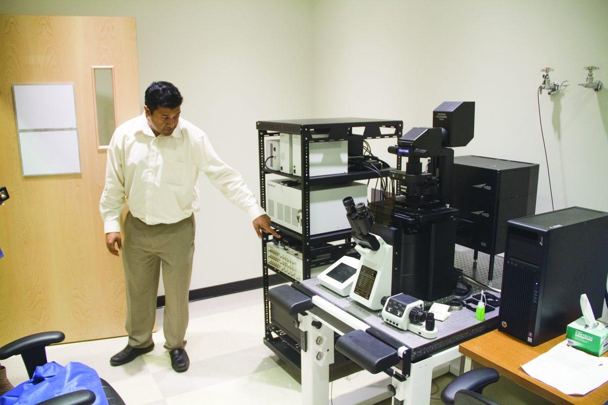
One year and nearly $3 million in the making, the Olympus Discovery Center provides new imaging systems expanding research opportunities for students, faculty and outside researchers.
The Center, housed in the basement of the Natural Science and Engineering Research Laboratory, allows researchers to utilize live cell imaging capabilities. This means students can administer medications to lab animals or inject substances into cells and see the results in real time, increasing the accuracy of the data.
Additionally, the equipment includes two confocal microscopes, one of which allows students to view fluorescent molecules and emissions using a laser, while the other allows students to perform high-speed and high-resolution imaging through a spinning disk. Students can also observe molecular interactions using total internal reflectance fluorescence, which allows small regions of compounds to be viewed by exciting its surface molecules.
Director of Core Research Facilities Jimmy Gowrisanker said he hopes the time and money put into the Center will pay off in the long run, and believes the lab is well-equipped to do that.
“One of my goals is for this lab to be set up in such a way that it’s marketed outside the university as well,” he said. “Hopefully this will foster cross-university collaboration, which can bring in money that will be put back into the Center.”
Olympus representatives teach two to three hour group training sessions for those wanting to use the lab, with instructions on how to properly use the equipment. This gives faculty time to focus on their research and allows students to begin working faster. In addition to the training, researchers also have to pay a fee averaging $24 per hour for equipment use.
UTD alumna Grishma Pradhan uses the Center for her ongoing research and said she is thankful for her training, which helped her understand not only the equipment, but the software behind it.
“Since we have representatives who are already here and we have them on call, if we don’t feel comfortable using the equipment after getting trained, we can set up one-on-ones with them, so they can make sure you know how to use it properly,” she said.
Pradhan, who works with live animals, said she is excited about the added depth the Center will bring to her research, especially due to its high-tech nature, including a multiphoton imaging system, which is one of only two in the country.
“We apply medications to animals all the time, but we don’t actually see what is going on in the body,” she said. “With the new equipment, we can see a live feed of what’s going on.”
Neuroscience junior Adil Ahmed, said he is looking forward to using the new equipment.
“We can do a lot of things that we couldn’t do before, like live-cell imaging,” Ahmed said. “I feel like it’s going to be helpful and provide opportunities for everyone in the building.”
Pradhan said with the Center’s advanced technology and ongoing studies, it is already helping students conduct experiments they previously weren’t able to.
“Before, we had all of these ideas, but we couldn’t do them because we couldn’t analyze or image them,” Pradhan said. “But now, with the new microscopes, there’s so many things we can do. We can better analyze our results and look at them in different ways.”


